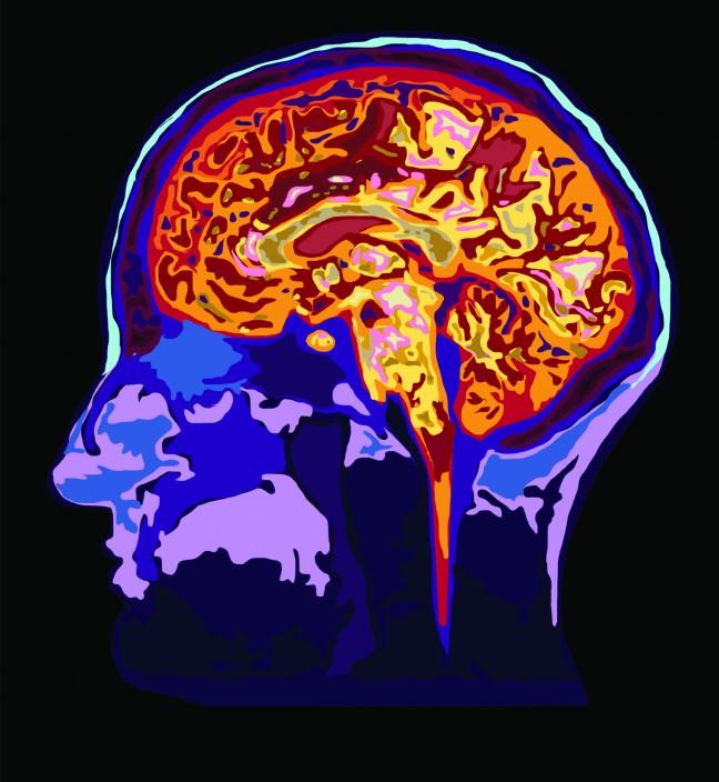
A team of researchers at University of Wisconsin has developed a noninvasive method to track brain development in children using two different brain imaging techniques.
In collaboration with researchers from Brown University and King’s College-London, UW researchers Doug Dean, Brittany Travers, Nagesh Adluru and Andrew Alexander studied myelination, a process through which the myelin sheath develops in the brain. Myelin facilitates transmission of nerve impulses and also protects neurons in the brain, Dean said.
Dean said there is very little myelin present in the young brain, but as people grow, the amount increases. Scientists have assumed that myelination is related to behavior and cognition development, he said. But they still do not understand how exactly these two processes are related.
The study examined how the myelin g-ratio, which is a ratio between the axon’s diameter and the myelin sheath’s diameter in the neuron, could be measured in children’s brains through two noninvasive techniques of magnetic resonance imaging.
Dean said measuring this ratio could help determine how quickly myelination occurs in children’s brains and possibly shed light on how myelination and behavior and cognition development are related.
The first technique, called DTI, or diffusor tensor imaging, provides detailed images of nerve cells and brain structures, but is unable to measure the g-ratio, Dean said. The second technique, called mcDESPOT, or multicomponent driven equilibrium single pulse observation of T1 and T2, measures the amount of myelin, but does not provide images of the whole neuron and brain structure. Both of these combined have allowed researchers to measure the g-ratio and connect it to other brain structures, he said.
“This has been some very interesting work about some properties of myelin g-ratio and its [relation to] optimal signal transmission in the brain,” Dean said.
According to the study’s findings, there is a specific g-ratio that facilitates optimal behavior and cognition development in the brain. In young children, this ratio starts around one and then decreases to 0.6-0.7 as the brain develops.
Dean said the study’s findings open up avenues for future research of brain development. He said potential areas of future research could involve studying the g-ratio in animal brains or through a microscope instead of an MRI.
Travers said the ratio could also have implications for future research of developmental disorders like autism. She said in most cases, it is difficult to point to a particular cause of a disorder in the brain, but the study’s techniques could shed some light on that.
“This is a really exciting study and would give us some more specifics to better understand what exactly is happening in myelin development in the brain,” Travers said.


