Studies on the retina at Sinha lab at the University of Wisconsin could provide valuable insight for blindness treatment, UW senior and Undergraduate Research Intern Paul Derr said.
Raunak Sinha, who is the principal investigator at the Sinha Lab, said macular degeneration is the most common form of blindness and other diseases causing blindness also come with major living impairments.
“People can’t see and it’s debilitating. It has a huge impact on their everyday life,” Sinha said.
According to Sinha, a large number of research efforts focus on restoring vision using stem cells. Labs are trying to create retinas using stem cells, but they lack the knowledge of how downstream photoreceptors must behave to make the neural circuits functional.
The retina is the neural tissue in the back of the eye converting photons of light into electrical signals and sends them to the brain. The visual pathway is responsible for 80% of the sensory input in the brain, Derr said.
“Overall, vision is just this preeminent domineering force for our interpretation and our understanding of the world,” Derr said.
Most blindness occurs because of the loss of photoreceptors at the top of neural circuits in the retina and most treatments for blindness use downstream circuitry to restore vision, Derr said. His work is exploring if these circuits can still be functional without the presence of upstream photoreceptors.
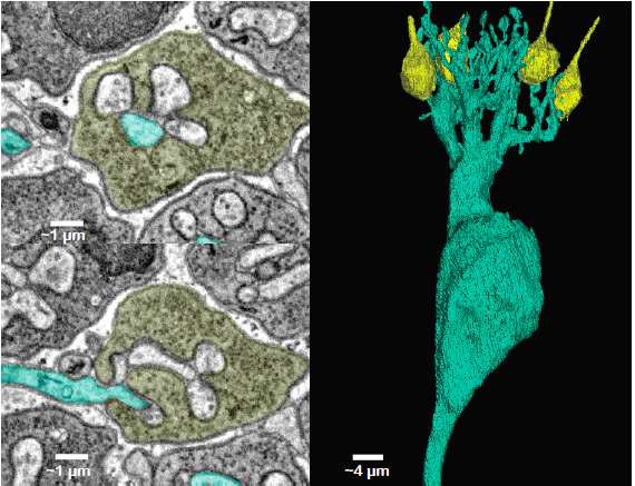
Derr uses a technique called single cell serial block face electron microscopy to observe cells in neural circuits. He said this technique takes an image of a tissue, slices off a thin layer of the tissue, then repeats this process to create a three dimensional image of the tissue.
Derr was investigating if having the proper morphology in the neural circuit structure — or connection of neurons — contributed to the system, or if lacking the proper functionality in the structure made the system entirely inoperable. What he found was encouraging.
“Just having the structure alone, without any functional contribution, was still enough to majorly improve the system,” Derr said.

Derr is observing three different models of the retina — a fully functioning and healthy model, a second one which is completely dysfunctional and inoperable and a third model which is functionally inoperable but with a proper structure.
Lamination is the quality in the retina making the location of a neuron determine its functional properties, Derr said. This allows researchers to draw expectations about how it should operate.
“The retina is a very beautiful neural circuit, in the sense that it’s very well organized,” Sinha said.
Derr’s project found in the entirely dysfunctional model of the retina, the neurons had complete disarray in the lamination, but the model with just the proper structure had proper lamination.
Derr won the sophomore research fellowship for this project —which was conducted with the retina of mice — his next project will use primate retina.
“Primates are kind of the gold standard for not only retinal work, but most neurobiology,” Derr said.
Non-human primates are much closer to humans than mice are from an evolutionary perspective, Derr said. This is because of the presence of the fovea, an anatomical specialization found only in primates.
The fovea is a concentrated area of high photon convergence in the center of the eye and gives humans high acuity color vision, which is why humans can see text and recognize faces.
“Anything you’re looking at, you’re looking at it with your fovea,” Derr said. “With that understanding, we have to realize that everything in our eyes is shaped to accentuate and support the fovea.”
The presence of the fovea results in non-homogenous vision, Sinha said, so eyes have high spatial recognition in the center while the peripheral vision does not. Conversely, the fovea cannot detect speed of motion as well as the peripheral.
The work in the Sinha lab is conducted in a dark room to present visual stimuli to models without any outside interference, Sinha said. The models they use are living tissues extracted and placed under a microscope to examine its response to stimuli.
“Our experiments are very rewarding because you get to see a neuron in action,” Sinha said.
Researchers are able to take stem cells from patients suffering from blindness due to gene mutations and create a model of the patient’s retina, then these models can be used for drug testing, Sinha added. The lab uses its knowledge of downstream circuitry and techniques to analyze the efficacy of certain drugs on these models.
Sinha said the goal of their research is to look at fundamental questions of the fovea and understand it from different perspectives. This could supply the necessary background for future treatments of blindness.
“This is the most important part of primate retina and yet we don’t know much about its signaling,” Sinha said. “We are only beginning to understand its uniqueness and the specializations involved.”


