


A study from researchers at the University of Wisconsin Carbone Cancer Center has laid out a new way to monitor the progress and success of cancer treatment practices.
The study was done in response to flaws in immunotherapy practices, which use parts of the immune system to fight diseases like cancer. Christian Capitini, Carbone Cancer Center member and pediatrics assistant professor at the UW School of Medicine and Public Health, authored the study.
“When we inject cell therapies in cancer, we don’t really have a good way of tracking cells after they’re injected,” Capitini said.
Capitini’s team experimented with MRI techniques to track “natural killer” cells after they were injected with fluorine-19, a non-radioactive, naturally occurring isotope. Natural killer cells, which are key players in immunotherapy, appear in around 5 percent of people’s peripheral blood cells, he said.
“They basically go around the body and look for virally infected cells, and it’s thought that they kill spontaneous development of cancer,” Capitini said.
Currently, most methods used to track cells involve radioisotopes because that method is more sensitive. While special MRI techniques can pick up fluorine-19, radioisotopes are identified with nuclear imaging, according to Sean Fain, study co-author, Carbone Cancer Center member and medical physics professor.
To pick up a signal with nuclear imaging, around 1,000 to 2,000 cells need to be traceable. For an MRI to track cells labeled with flourine, there must be around 10,000 to 30,000 of them, Fain said.
But radioisotopes are not without their own problems. They typically decay in a number of hours, so the cell can’t be tracked for long periods of time using that method. Capitini said they’re also more difficult to locate.
“[With radioisotopes] you can see a big splotch of where your cells are, but you don’t know exactly what organ or what area they’re in; you just know in general where they are geographically,” Capitini said. “For example, if you have a tumor in your brain, with radioisotopes, you would see a signal in your head, but you wouldn’t know if it’s actually in the brain tumor or just in your brain. With an MRI scan, you’d be able to distinguish the two.”
Fain said another advantage of using MRI is that it doesn’t use radioisotopes, so it’s more attractive for patients who are sensitive to radiation risk, especially children.
Tracking these cells is important to help doctors understand why immunotherapy doesn’t work in some patients, Capitini said.
“Is it just because the cells don’t kill [the tumor]? Or is it because they can’t find it? The goal is to use MRI as a guide to help you figure out if they found the tumor or how long they’re surviving after you inject them,” he said.
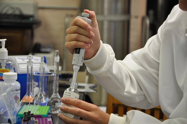
In their process, the team first took human natural killer cells from regular blood and grew them in a lab to create a larger population. Those cells were incubated with fluorine-19, which was later taken up by the cells. Then they put tubes of those cells in an MRI spectrometer that was specifically designed to detect fluorine atoms, according to the study.
After proving they could see the natural killer cells in an MRI machine, the next step was to test the method in a living organism.
The group used mice, which are a common starting point for many laboratory experiments. Immune-deficient mice were used in this study because their immune systems don’t reject human cells, Capitini said.
The team tried two methods: first, injecting natural killer cells directly into a tumor, and second, injecting intravenously. So far, direct injection has been the most successful, Fain said.
Unfortunately, this is the lesser-preferred method, he said. To do a direct injection, the tumor needs to be relatively close to the surface, and there are certain parts of the body — like the brain — where injecting directly can be risky.
“The use of imaging is pretty important to enable monitoring therapy and to establish whether certain approaches to immunotherapy are working better than others,” Fain said. “I think everyone in the field of cancer imaging is racing right now to try and provide a means to image cell trafficking, but it’s uncertain right now which technique will be preferred and whether we’ll get there.”